6 AI Tools That Support Veterinary Radiologists in Practice
Compare 6 AI tools for veterinary radiologists: SignalPET, Vetology, Radimal, Antech RapidRead, PicoxIA and Kairos, with human review kept in the loop.
 Eimantas Kazėnas Marketing & Tech
- Published
- 10 min read
On this page At a glance
Independent and reader-supported. Some links may earn us a commission; it never changes our rankings. Editorial policy
A veterinary radiologist’s work involves more than identifying findings on an image. It also includes reviewing clinical context, preparing clear reports, managing case volume and helping referring teams decide what to do next. AI tools can support parts of that workflow, but they differ in what they analyze and how much human review they include.
Some tools screen images and generate preliminary findings. Others help organize studies, draft reports from a radiologist’s observations or connect practices with specialist interpretation. For clinics and imaging groups, the practical question is how well each tool fits into the workflow and where the final clinical judgment remains.
At a glance
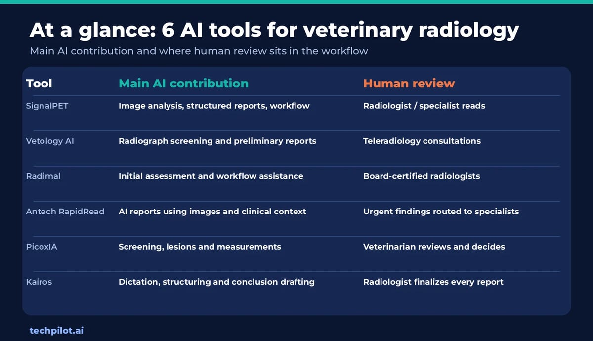
| Tool | Main AI contribution | Human review in the workflow |
|---|---|---|
| SignalPET | Image analysis, structured reports and radiology workflow support | Offers access to veterinary radiologists and specialist reads |
| Vetology AI | Radiograph screening and preliminary reports | Offers teleradiology consultations |
| Radimal | Initial image assessment and radiologist workflow assistance | Connects practices with board-certified radiologists |
| Antech RapidRead | AI-generated radiology reports using images and clinical context | Routes selected urgent findings for specialist review |
| PicoxIA | Radiograph screening, lesion identification and measurements | Veterinarians review findings and make clinical decisions |
| Kairos | Dictation, report structuring and conclusion drafting | Radiologist reviews, edits and finalizes every report |
What AI can and cannot do in veterinary radiology
AI tools can help make imaging workflows more consistent and timely. Depending on the product, they may flag patterns, measure selected structures, organize images or help turn a radiologist’s notes into a structured report. These functions can reduce repetitive work and draw attention to findings that merit closer review.
The tool’s role depends on what it receives and produces:
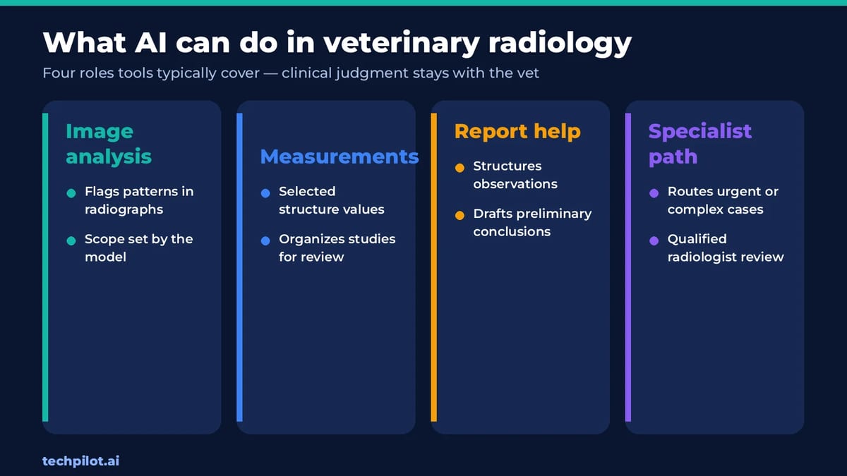
- Image analysis evaluates radiographs or other imaging data for patterns within the model’s intended scope.
- Measurement and image organization can support tasks such as calculating selected values or arranging studies for review.
- Report assistance can structure observations, draft language or generate preliminary conclusions.
- Specialist interpretation adds a qualified radiologist’s review when the case needs expert assessment.
AI output should be evaluated in the context of the case, including patient history, clinical signs and image quality. Veterinary radiology organizations emphasize the importance of keeping a veterinarian involved in interpreting AI output. The sections below describe the specific role each product can play in that process.
6 AI tools for veterinary radiology workflows
1. SignalPET
SignalPET combines AI-supported image analysis with reporting and access to veterinary radiology services. Its workflow includes an Immediate Report for rapid AI insights and more comprehensive reporting options that can use clinical context and a veterinarian’s specific questions. Practices can also submit cases for review by veterinary radiologists.
That range of support can help practices use AI as part of a broader imaging process. A case can begin with an AI review and move to a more detailed report or specialist interpretation when the clinical question calls for it. SignalPET also provides a cloud-based imaging environment that supports radiography and other imaging modalities, including ultrasound, MRI and CT.
SignalPET’s integrated workflow gives it a broad role in this comparison: AI insights, reporting options, imaging tools and radiologist access are presented as connected parts of the service. Teams can evaluate whether that combination reduces handoffs and helps them move from an image to an appropriate clinical next step.
For radiologists, the platform’s potential value is in connecting image review, reporting and escalation within one workflow. For referring practices, structured findings can support the medical record and conversations about next steps. The specific report and review path should match the case, since an AI screening result and a full diagnostic interpretation serve different purposes.
What to assess:
- Which report type fits routine, urgent and complex cases
- How clinical history and the referring veterinarian’s question inform the report
- How to submit a case for specialist review
- Which imaging modalities and existing systems the workflow supports
- How findings, reports and images are stored and shared
2. Vetology AI
Vetology AI analyzes veterinary radiographs and provides screening results or preliminary reports. The product is part of a wider teleradiology offering, so practices can use AI support alongside access to radiologist consultations. Vetology has also described a radiologist-facing preliminary tool for managing and working through cases.
For a radiologist, AI screening may provide an additional review point before a report is finalized. For a general practice team, a preliminary report can help organize the initial assessment while the veterinarian considers the patient history and clinical presentation. The tool is designed to support clinical review, rather than make the final decision about diagnosis or treatment.
Vetology has updated its AI classifier architecture and reports multiple classifiers for canine and feline imaging. Practices should confirm the current scope of the models, including which body regions, views and findings are covered. Performance on one type of radiograph does not automatically establish performance across all studies.
What to assess:
- Which species, views and findings are included in the current classifier set
- How AI screening results differ from a complete radiologist interpretation
- How to request a teleradiology consultation
- How reports fit into the practice’s image and medical-record workflow
- What validation information is available for the use cases you plan to deploy
3. Radimal
Radimal combines AI-supported image assessment with radiologist services and imaging workflow tools. Its Initial Assessment reviews X-rays and provides preliminary indications for the veterinary team to consider. The platform also offers a way to request interpretation from a board-certified veterinary radiologist.
Radimal includes tools for radiologist worklists, image viewing and report preparation. Its radiologist-facing materials describe AI support for tasks such as hanging protocols and generative report writing assistance, alongside the Initial Assessment feature. This means the product can be considered from both the clinic’s perspective and the perspective of imaging professionals managing cases.
The AI assessment is not a definitive diagnosis. The team should interpret it with the patient’s clinical information and request specialist review when needed. A practice should also confirm which features are available to its specific user type, since the workflow and tools may differ between referring clinics and radiologists.
What to assess:
- Which Initial Assessment findings are covered for your patient population
- How radiologist consultation is requested and delivered
- Which worklist, viewing and report-writing tools are included
- How the platform connects with existing imaging and practice systems
- How the team distinguishes preliminary AI findings from specialist conclusions
4. Antech RapidRead
Antech RapidRead generates AI-supported radiology reports for dogs and cats. The service considers radiographic findings alongside signalment and clinical observations provided with the case. Antech describes coverage for common findings in the thorax, abdomen and limbs, with reports designed to return within minutes.
The service also includes a human review pathway for selected emergent findings. Antech says certain findings are routed to a board-certified veterinary specialist for a STAT review, and practices can request additional interpretation through its imaging consultation services.
RapidRead’s workflow combines an AI report with access to specialist support. This can help teams decide when a case can proceed with the available information and when an expert read is needed. Teams should confirm the current scope, turnaround expectations and the difference between an AI-generated report and a radiologist consultation.
What to assess:
- Whether the body regions and findings cover the cases you receive
- What patient information and clinical observations are included
- Which findings trigger specialist review and what turnaround applies
- How to request additional radiologist input
- How RapidRead reports are incorporated into the medical record
5. PicoxIA
PicoxIA analyzes veterinary radiographs and provides a list of lesions identified by its algorithm. The product describes support for thoracic, abdominal and pelvic X-rays, including measurements such as vertebral heart size. This makes its role focused on image screening and selected measurements rather than the full radiology workflow.
An automated result can give a veterinarian or radiologist another structured point of reference during image review. The clinician still needs to assess whether the result fits the image, patient history and other findings. Teams should pay attention to the specific imaging views and conditions the product is designed to analyze.
PicoxIA also describes integration with image acquisition software. Integration may reduce the steps needed to submit or review a study, but the details depend on the equipment and software already used by the practice. Confirm the workflow before expecting it to work as a direct extension of the existing imaging setup.
What to assess:
- Which species, body regions and radiographic views are covered
- Which lesions and measurements the product reports
- How AI results are displayed alongside the original radiographs
- Whether your acquisition software is supported
- What validation evidence applies to your intended clinical use
6. Kairos
Kairos supports veterinary radiologists during report creation. It uses AI-assisted dictation and generative reporting to turn a radiologist’s findings into structured conclusions and recommendations. Unlike image-screening tools, Kairos does not interpret the radiographs themselves. The radiologist supplies the findings, then reviews and edits the generated report.
This focus addresses a different part of radiology work: drafting, formatting and maintaining consistency across reports. Radiologists can dictate or enter observations and use the platform to generate a structured conclusion. Templates and preferences can help align the output with an individual’s reporting style.
Kairos is described as an open beta, and its model transparency information notes that it is undergoing validation. Its output may require corrections, particularly in complex or unusual cases. That makes human review essential before a report is finalized or shared with a referring veterinarian.
What to assess:
- Whether dictation and report generation match your existing reporting style
- How templates, tone and formatting preferences are configured
- How the tool handles complex cases and unusual modality or species combinations
- What privacy and data-handling terms apply to report content
- How beta status and validation plans affect clinical use
How to evaluate AI tools for veterinary radiology
A product description can show what a tool is designed to do, but a practice needs to know how it performs on its own cases. Start with a defined set of use cases and agree on how the team will judge the output.
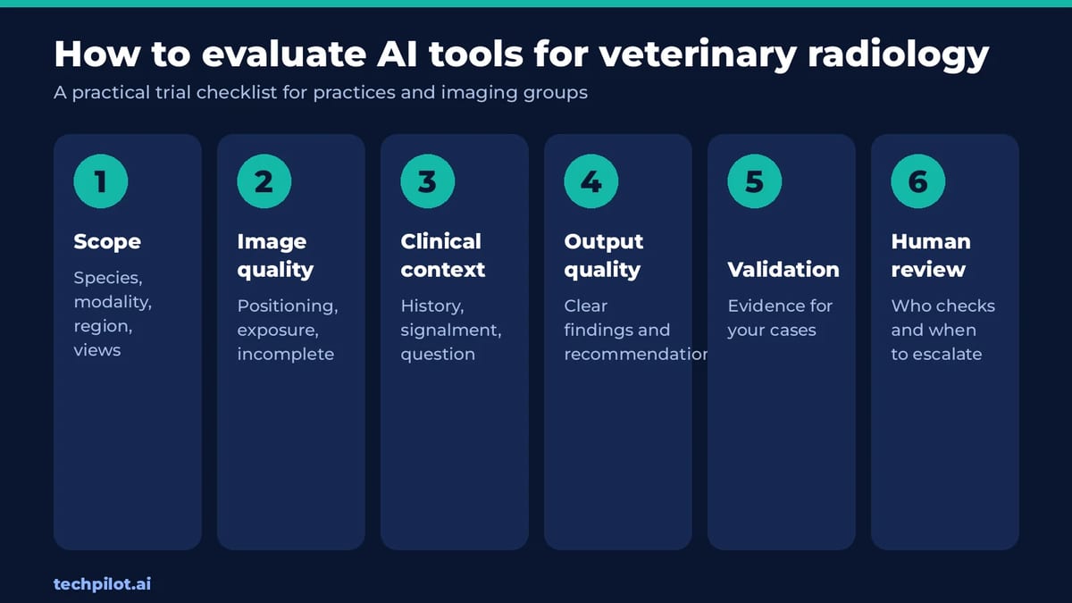
A practical evaluation can include:
- Scope: Confirm the species, modality, body region, views and findings supported.
- Image quality: Test how the system handles positioning, exposure and incomplete studies.
- Clinical context: Check whether the tool accepts history, signalment and the referring question.
- Output quality: Review whether findings, measurements and recommendations are clear and usable.
- Validation: Request evidence for the tasks and patient population relevant to your practice.
- Human review: Define who checks the AI output and when to request specialist interpretation.
Use a representative sample of normal, routine, urgent and complex studies. Compare AI output with radiologist interpretations, then document where the tool agrees, misses a finding or produces a result that needs editing. This gives the practice a more useful basis for deciding where AI can fit.
FAQs about AI tools for veterinary radiologists
Can AI replace a veterinary radiologist?
AI can support image review, measurements, triage or report drafting, but it does not replace a radiologist’s clinical judgment. Each tool has a defined scope and may miss findings or produce output that needs correction. Practices should set clear review and escalation steps, especially for urgent, complex or unexpected cases.
Do all veterinary radiology AI tools analyze images?
No. Some products evaluate radiographs and return screening findings or measurements. Others work from observations entered or dictated by a radiologist and help draft the report. Confirm what information the tool receives and whether it interprets images, supports reporting, or performs both functions before comparing it with another product.
What should a practice review during an AI trial?
Test representative cases across species, body regions, image quality and clinical urgency. Compare AI output with the final interpretation, and record missed findings, unclear language and editing time. Also check integration with the imaging workflow and how quickly the team can request specialist review when the case needs additional expertise.
How should a team handle AI findings that do not match the clinical picture?
The team should review the original images, patient history and clinical signs, then follow its established diagnostic process. If the discrepancy remains important, seek a qualified radiologist’s interpretation. Document how the concern was resolved. AI output should not override clinical judgment or become the sole basis for a treatment decision.
What is the difference between an AI report and a radiologist report?
An AI report is generated by a model within a defined scope. A radiologist report is prepared or reviewed by a qualified specialist who can assess the images in context and apply professional judgment. Some services offer both, so teams should confirm which report they received and whether a human reviewed it.
Found this useful? Share it with someone comparing AI tools.